
When your eye doctor says you need an imaging test, it’s not just a routine check-it’s a window into the health of your retina, optic nerve, and blood vessels behind your eye. Three tools dominate modern eye care: OCT, fundus photography, and fluorescein angiography. Each gives a different kind of information. Together, they help catch problems like diabetic retinopathy, macular degeneration, and rare retinal diseases before they steal your vision.
What OCT Really Shows
Optical Coherence Tomography (OCT) is like a high-resolution ultrasound for your eye. It doesn’t use light to take a picture like a camera. Instead, it uses infrared light to scan layer by layer, building a 3D map of your retina. Think of it as slicing open your eye in a digital lab, seeing every microscopic structure without touching it. Spectral-domain OCT (SD-OCT), the most common type today, captures images with a resolution of 5 to 7 micrometers. That’s thinner than a human hair. It shows fluid buildup, swelling, thinning of layers, and even tiny holes in the macula. For someone with diabetic eye disease, OCT can reveal fluid trapped in the retina long before they notice blurry vision. In age-related macular degeneration (AMD), it shows whether the disease is dry (thinning) or wet (fluid leakage under the retina). Newer swept-source OCT (SS-OCT) goes deeper. It can see into the choroid-the layer of blood vessels under the retina that feeds the retina. This matters because many diseases, like punctate inner choroidopathy (PIC), start there. SS-OCT scans 100,000 to 400,000 lines per second, compared to 20,000-85,000 for older models. Faster scans mean less blur from blinking or eye movement.Fundus Photography: The Classic Snapshot
Fundus photography is the oldest of the three. It’s what most people picture when they think of an eye exam: a bright flash, a wide-angle photo of the back of the eye. Cameras like the Zeiss FF 450+ capture detailed images of the optic nerve, retina, and blood vessels. These photos are used to track changes over time. Why keep taking the same picture year after year? Because small changes matter. A new blood vessel growing near the optic nerve, a tiny hemorrhage near the macula, or a subtle shift in the optic nerve’s color can signal early diabetic retinopathy or glaucoma. Unlike OCT, fundus photos show the big picture-what’s happening across the whole retina, not just one slice. But fundus photos have limits. They can’t show fluid inside layers. They can’t tell if a blood vessel is leaking. They’re also affected by cataracts, pupil size, or poor focus. If your eye is cloudy or your pupil is small, the image quality drops. That’s why it’s rarely used alone anymore.Fluorescein Angiography: The Dye Test
Fluorescein angiography (FA) is the only one that involves a shot. A yellow dye called fluorescein is injected into a vein in your arm. As it travels through your bloodstream, a special camera takes rapid photos of your retina. The dye lights up blood vessels, making leaks, blockages, and abnormal growths glow under blue light. It’s still the gold standard for spotting leaking vessels in diabetic macular edema. Studies show FA detects leakage with 100% sensitivity, while SD-OCT only catches 79%. That 21% gap? That’s where patients might miss treatment. FA also shows areas where blood flow is blocked-called non-perfusion zones-which can predict future vision loss. But FA isn’t perfect. It takes 10 to 30 minutes. You might feel nauseous or get a yellow tint to your skin. Rarely, people have allergic reactions. It can’t show the deeper layers of the retina. And because it’s a 2D image, it’s hard to tell exactly where the problem is in 3D space.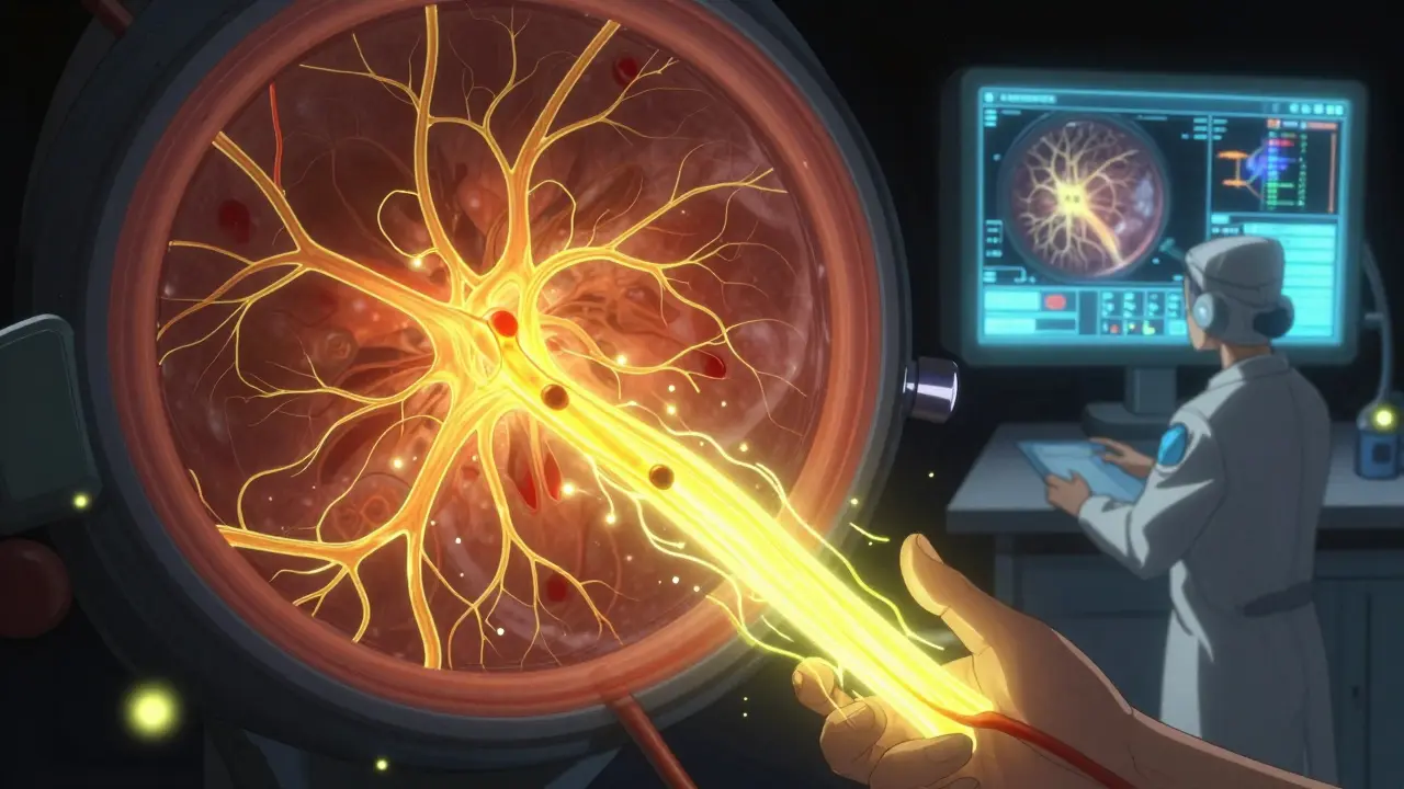
OCT Angiography: The Game-Changer
OCT angiography (OCTA) is the newest kid on the block. It doesn’t need dye. It uses the same OCT machine but with smarter software that detects blood flow by tracking movement between scans. It gives you a 3D map of retinal and choroidal blood vessels-without a needle. This matters because it shows details FA can’t. OCTA can separate the capillary layers: superficial, middle, and deep. In diabetic retinopathy, it can show tiny areas of capillary dropout that FA might miss. In Coats disease, it reveals abnormal vessels growing in layers you can’t see with traditional imaging. A 2023 study found that swept-source OCTA detected 57% more retinal capillary hemangiomas than spectral-domain OCTA. It also shows wider areas of the retina-up to 12mm across-compared to FA’s 30-degree view. That means it can catch problems near the edge of the retina that older methods overlook. But OCTA has its own problems. If you move your eye even a little, the image blurs. Patients with poor vision, tremors, or children often can’t hold still long enough. It also can’t show dye leakage, which is critical in some cases. So while it’s amazing for mapping blood vessels, it doesn’t replace FA entirely.How They Work Together
No single test tells the whole story. That’s why clinics use multimodal imaging:- For diabetic retinopathy: OCT shows fluid swelling; FA shows leaking vessels; OCTA shows early capillary loss.
- For macular degeneration: OCT confirms drusen and fluid; FA identifies abnormal blood vessels; OCTA maps their growth pattern.
- For Coats disease: Fundus photos show white, fluffy exudates; OCT reveals fluid pockets and cholesterol crystals; OCTA shows abnormal vessel networks.
- For PIC: OCTA finds tiny choroidal non-perfusion areas invisible to FA or fundus photos.
What You Can Expect During Testing
- OCT: You sit in front of a machine, rest your chin, stare at a light. No contact. No light flash. Takes 5-10 minutes. You might hear a clicking noise.What’s Changing Right Now
The field is moving fast. New OCTA systems, like the Spectralis platform, have improved algorithms that reduce motion blur and show deeper choroidal vessels. Wide-field OCTA now covers up to 12mm of the retina-close to what FA does, but without dye. Artificial intelligence is starting to analyze these images automatically. It can count microaneurysms, measure capillary density, or flag early signs of disease before a doctor even sees them. This isn’t science fiction-it’s already in clinics in London, Boston, and Sydney. The big question isn’t whether OCTA will replace FA. It’s when. For some conditions, like macular edema from vein blockages, FA is still irreplaceable. But for monitoring progression, tracking vascular changes, or screening high-risk patients, OCTA is becoming the first choice.What You Should Know
- OCT and OCTA are painless. No side effects.If you have diabetes, high blood pressure, or a family history of eye disease, these tests aren’t optional. They’re your best defense against silent vision loss. The sooner you catch a problem, the more you can save.
Is OCT the same as a retinal scan?
Yes, OCT is a type of retinal scan, but not all retinal scans are OCT. Fundus photography is also a retinal scan, but it takes 2D color photos. OCT takes 3D cross-sections. Think of OCT as a detailed MRI of the retina, while fundus photos are like a high-res camera snapshot.
Do I need to have my pupils dilated for OCT or OCTA?
Not always. Many modern OCT machines can capture good images even with small pupils. But for the clearest view-especially if your doctor suspects deep disease like choroidal problems-dilation is still recommended. It’s not required for every visit, but it helps when you’re being evaluated for serious conditions.
Can OCTA replace fluorescein angiography completely?
Not yet. OCTA is excellent for mapping blood vessels and detecting blockages, but it can’t show dye leakage-which is critical in diabetic macular edema, retinal vein occlusions, or some forms of macular degeneration. FA still wins in those cases. Many clinics now use OCTA first, then do FA only if needed.
Why does my doctor order both fundus photos and OCT?
Because they show different things. Fundus photos show the big picture: where blood vessels are, if there’s bleeding or scarring. OCT shows the layers beneath: is there fluid? Is the retina thinning? Is there a hole? Together, they give a complete picture. One without the other is like having only half the story.
How often should I get these tests if I have diabetes?
If you have diabetes, you should have at least one comprehensive eye exam with OCT and fundus photography every year. If you already have retinopathy, your doctor may recommend scans every 3-6 months. The frequency depends on how advanced the disease is and how well your blood sugar is controlled. Don’t skip them-even if your vision feels fine.
If you’ve been told you need one of these tests, don’t worry. They’re routine, safe, and often the only way to catch problems before they become serious. The goal isn’t to scare you-it’s to protect your sight, one scan at a time.


Tom Forwood February 8, 2026
Just had my first OCTA last week-no dye, no fuss. Felt like a sci-fi movie where they scan your brain except it’s your eye. The tech is wild. My doc showed me the capillary layers like they were a roadmap to my retina. I swear I saw a tiny dead end that looked like a cul-de-sac. Weirdly satisfying.
And yeah, no dilation. My pupils were tiny, and it still worked. Modern medicine is kinda magical when it doesn’t hurt.
Also, why does everyone still call it a ‘retinal scan’? It’s not one thing. It’s a whole damn toolkit. We need better vocabulary.
Alex Ogle February 8, 2026
Man, I’ve been through all three. Fundus photo? Bright flash, feels like someone shined a flashlight up your soul. FA? That warm dye shot? Feels like your arm’s on fire for a second, then you’re yellow for hours. My wife thought I was jaundiced.
OCT? Zero drama. Just sit there, stare at a light, listen to the machine click like a dying robot. I’ve had it five times in two years. My diabetes’s been stable, but they catch the tiniest fluid shifts. Like, I didn’t even know I had swelling until the scan showed it.
And OCTA? That’s the future. No needle, full 3D blood map. I’m convinced they’re going to replace FA entirely in five years. Just need better software to stop the motion blur. My dog moved my head once. Scan was garbage.
Bottom line: don’t skip the scans. Even if your vision’s perfect. Your retina doesn’t scream until it’s too late.
Simon Critchley February 9, 2026
As a guy who’s read 17 papers on multimodal retinal imaging, let me drop some jargon: OCTA’s sensitivity for microvascular dropout is 92% (CI 88–95%) vs. FA’s 79% for leakage detection. But here’s the kicker-FA still holds the crown for quantifying non-perfusion zones because of its dynamic contrast kinetics.
OCTA’s spatial resolution? 5μm lateral, 7μm axial. SS-OCTA pushes choroidal penetration to 300μm. That’s game-changing for PIC and choroidal neovascularization. But motion artifacts? Still a bitch. Especially with nystagmus or Parkinsonian tremors.
And don’t get me started on AI segmentation algorithms. The 2023 JAMA Ophthalmology meta-analysis showed a 31% reduction in false positives with deep learning-based capillary mapping. We’re not just imaging anymore-we’re predicting.
TL;DR: FA isn’t dead. It’s just sharing the throne.
Chelsea Cook February 10, 2026
So let me get this straight: we’re doing a full-on sci-fi eye scan on diabetics, but some of us still think ‘dilation is optional’? 😂
My mom skipped her FA because ‘it’s too much hassle.’ Two years later, she lost 30% of her peripheral vision. Turns out the leakage was hiding under a cataract. Fundus photo looked fine. OCT showed nothing. FA? Full-on fireworks.
Stop romanticizing ‘painless tech.’ Sometimes the pain is the price of sight. And yes, I’m still mad at my old doctor for not pushing harder.
Joseph Charles Colin February 11, 2026
Clarifying a common misconception: OCT does not use infrared light to ‘scan layer by layer’ in the way ultrasound does. It relies on interferometry-measuring the interference pattern of low-coherence light reflected from retinal layers. The axial resolution is determined by the source bandwidth, not depth penetration.
SS-OCT’s advantage isn’t just speed-it’s the 1050nm center wavelength, which reduces scattering in pigmented tissues and enhances choroidal signal. This is why it outperforms SD-OCT in detecting choroidal thickening in PIC or pachychoroid syndromes.
Also, OCTA’s capillary layer segmentation is algorithm-dependent. Most commercial systems use a ‘projection-resolved’ method, but newer deep learning models (e.g., U-Net variants) now enable true volumetric flow mapping. This is why 2023 studies show 57% more hemangiomas detected.
Brandon Osborne February 11, 2026
Ugh. Another tech brochure masquerading as medical advice. You people act like OCTA is some miracle cure, but it’s just a fancy camera with a software filter. Meanwhile, real doctors are still using FA because it WORKS.
And let’s talk about that ‘AI analysis’ nonsense. You think some algorithm trained on 500 scans can catch what a trained ophthalmologist sees? Please. I’ve seen AI flag normal vessels as ‘abnormal’ and miss actual neovascularization because the lighting was off.
Stop outsourcing your brain to machines. The eye is not a smartphone. You don’t ‘update’ vision loss with an app.
John McDonald February 13, 2026
Love this breakdown. I’m a nurse who’s helped run these tests for 12 years. I’ve seen patients cry because FA made them nauseous. I’ve seen others laugh because OCTA was so easy they thought it was a game.
One thing I always say: don’t fear the dye. The worst part is the yellow skin. It’s temporary. The vision you save? Not.
And if you’re diabetic? Get the full combo every year. Even if your HbA1c is perfect. The retina doesn’t care about your diet-it cares about blood flow. And that’s what these scans track.
Also, shoutout to the techs who hold your hand through this. They’re the real heroes. No one remembers them. But they’re the ones making sure the images aren’t blurry because you blinked.
John Watts February 13, 2026
As someone who moved from Nigeria to the States, I’ve seen both sides. Back home, most clinics still use old fundus cameras-no OCT, no FA. People lose vision and don’t know why until it’s too late.
Here? We’ve got AI analyzing capillary density like it’s a weather map. It’s insane. But the gap is real. I’ve told 3 family members back home to save up, come here, get scanned. One of them had early diabetic retinopathy. Caught it. Now she’s on meds. Vision intact.
Don’t take this for granted. This tech? It’s a gift. Use it. And if you’re lucky enough to have access? Help someone who doesn’t.
Chima Ifeanyi February 15, 2026
Let’s be real. This whole ‘multimodal imaging’ thing is just Big Ophthalmology’s way of selling more scans. Who’s paying for this? Insurance? Medicare? You think your doc genuinely cares about your choroidal perfusion-or are they just hitting their quarterly imaging quota?
And OCTA? No dye? Sure. But it’s also 3x more expensive than FA. And 80% of the time, it doesn’t change the treatment plan.
Real talk: if you’re diabetic and your vision is fine, you don’t need a 3D blood map. You need better glucose control. Not a $600 scan.
Stop being manipulated by tech hype.
Elan Ricarte February 16, 2026
Bro. OCTA is the new Tesla. Everyone’s obsessed with it. But guess what? Most retinal diseases still get diagnosed with FA. Because the dye leaks. And that’s the whole damn point.
I’ve seen OCTA show ‘perfect’ vessels… then FA shows a full-on storm of leakage. You think a machine that tracks movement can catch a slow, sneaky leak? Nah.
Also, AI? Cool. But I’ve seen it miss a tiny hemorrhage because the image was ‘too noisy.’ Meanwhile, my grandma’s doc caught it with a magnifying glass and 30 years of experience.
Tech is a tool. Not a prophet.
Andrew Jackson February 18, 2026
It is with profound concern that I observe the uncritical embrace of technological overreach in medical diagnostics. The human eye, a divine instrument of perception, is being reduced to a data stream-a pixelated abstraction, parsed by algorithms written in Silicon Valley.
Who authorized this? Who gave the FDA the right to replace the physician’s judgment with machine learning models trained on datasets of unknown provenance?
Fluorescein angiography, a method refined over decades of clinical wisdom, is being sidelined in favor of a non-invasive, yet fundamentally incomplete, optical method. This is not progress. This is hubris.
And let us not forget: the body does not exist in 3D layers. It exists in divine order. To dissect it into spectral bands and flow maps is to violate the integrity of natural form.
We must return to the art of the clinical exam. The gaze. The history. The touch. Not the machine.
Kathryn Lenn February 19, 2026
Okay, but what if the machines are lying?
I read a study once where OCTA flagged 12% of healthy people as having ‘early diabetic retinopathy’ because of a software glitch. Then the FDA approved a new algorithm. Then they changed it again. Then they changed it back.
And now we’re supposed to trust it? What if the ‘dye’ in FA is just a placebo? What if the ‘fluid’ on OCT is just shadows? What if all this is just a way to keep people coming back for more scans so the clinics can keep billing?
My cousin got 7 scans in 6 months. He’s not diabetic. He has glaucoma. Why? Because ‘we need baseline data.’
Who’s really getting rich here? Not me. Not you. Definitely not the patient.